ADS

9 Nursing Care Plan for Personality Disorders

Personality is the traits and attitudes that can be suspected in someone and affect the patterns of cognitive, affective and behavioral. The patterns in a long time, consciously or unconsciously, and affect people's response and adaptation to the environment. Adaptation is the center of personality theory.


Definition of Personality Disorders

Kerberg (1984): maladaptive attitudes that produce or influence the psychological considerations, causing emotional disturbance and damage relations with others.

Maladaptive responses such as inflexible, rigid, is a pathological condition that causes the client stress, anxiety and depression.


1. Paranoid Personality Disorder

Nursing Care Plan for Paranoid Personality Disorder
Pervasive suspicion and unwarranted and not trust to others.
  • Often feel like to be kept others from feeling threatened or attacks from others.
  • Mood them sensitive and hostile, aloof.
  • Difficult to build a trusting relationship
  • Be careful in dealing with other people.
  • Pathological jealousy.
  • Inability to relax, lacking a sense of humor.
  • Critical to the others, but it is difficult to accept criticism.
Interventions:
  • Approach calm manner, empathy and avoid things that make jealous.
  • Due to suspicious clients will harm everyone, then the nurse must consider verbal and non-verbal signs to determine if the client is being aggressive or hostile.
  • All actions will be discussed first so he understands the reason and purpose, so as not suspicious.
  • Try to act according to the agreements made​​.
  • Support adaptive behaviors such as trust others are not always going to threaten, that self safe. Able to adapt to stressors.

2. Schizotypal and Schizoid Personality Disorder

Nursing Care Plan for Schizotypal and Schizoid Personality Disorder
The inability to form close personal relationships
  • This person is difficult to relate to others, and difficult to maintain intimate relationships.
  • Tend could develop into schizophrenia, and there is a genetic element.
  • The signs are: isolation, limited interaction with peers, social anxiety, school performance is not good, hypersensitivity, limitations in speech and thought process, no warmth / softness.
  • Do not care about the praise / criticism of others.
  • Affect blunt, flat, pulled away and looked cool.

Intervention:
  • Approach client calmly.
  • Adjust the level of talks with the relationship with the client, observe the verbal and nonverbal.
  • Include in group therapy.
  • Arrange level client relationships, for example, starting from the nurse-client relationship, the client - the client, the client with another group and finally group therapy.

3. Antisocial Personality Disorder

Nursing Care Plan for Antisocial Personality Disorder
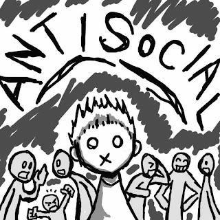
The person does not behave according to social norms.
Characteristics: failure to learn from experience, often acting risky and impulsive, no guilt and recurrent, exploit others, does not respect the rights of others, there is no loyalty and honesty.
  • Tend to be drug addicts.
  • They are manipulative and are happy to argue
  • Teams often fail to address the health because they often do unlawful acts and norms, cheat, do not have a plan as impulsive, there is a history of fighting, insulting and aggressive, could endanger themselves and others, failed to manage finances.

Interventions:
  • Be an example of how to interact with sound.
  • Do not get stuck client behaviors such as arguing, make sure the rules and sanctions that will be earned if not obedient, discuss the impact of his behavior over the years.
  • Include in group therapy, family and health care team work together so that all the same rules and be consistent.
  • Encourage clients to get along with others so that good self-esteem.


4. Borderline Personality Disorder
Nursing Care Plan for Borderline Personality Disorder
An instability in interpersonal relationships, self-image and feeling natural.
  • This guy has an ego that is not integrated and fragile.
Characteristics: do not have a clear identity, tend to use coping children, it is difficult to accept the fact.
  • When stressed, he will regression, spliting (looking at the world as a dichotomous, good - bad, positive-negative), deny and projections.
  • Tend to feel lonely, hurting himself.

Interventions:
  • Soothing at a critical time.
  • Because nurses often refer to either (when his will be obeyed) or bitchy sisters, it is often a conflict. So the nurse to avoid and resolve conflicts.
  • Create a list of daily activities, regulations, responsibility and a clear agreement.
  • Avoid self-harm acts directly or indirectly because he felt himself evil.
  • If necessary collaborative physician for appropriate medication symptoms.
  • Include in group therapy.


5. Histrionic Personality Disorder

Nursing Care Plan for Histrionic Personality Disorder
The behavior of clients ranging from high ego functions (attention-seeking) to the lower ego functions (want to have intercourse with anyone, impulsivity, and psychosis).
Behavior seen:
  • Feeling uncomfortable in a situation where he is not the center of attention.
  • Having sexual intercourse with many people.
  • Perform physical attractiveness to seek attention.
  • Diction made as attractive as possible.
  • Behave dramatic, as in the theater at the time of expressing his emotions.
  • Easy on the suggestion.
  • Like intimate relationship with anyone, even if other people do not feel it.
  • Overreaction to minor events
  • Arrogant and demanding behavior

This disorder in women, relationships with others are usually superficial, flamboyant, and prefer to rely on therapists
  • His ego makes him fantasize, fall in love with the therapist.
  • Coping: dissociation, repression.
  • Other maladaptive behavior: temper tantrums, manipulative, relentless demand.

Interventions:
  • Consistent, understanding, overcoming feelings of love or hate the therapist
  • Making the environment do not cause maladaptive behavior.
  • Discuss about fantasy, brought to reality
  • Teach healthy behaviors in achieving goals, teach constructive coping.
  • Taught the principles and healthy behaviors.

6. Narcissism Personality Disorder
Nursing Care Plan for Narcissism Personality Disorder
People who are enjoying themselves and are always acting to please him.
  • The person is able to work but are often asked for help because of difficulty in dealing closely with other people, no neurotic symptoms, sexual difficulties, and there is a chronic feeling of emptiness.

Characteristics:
  • Want to be considered superior.
  • There is a fantasy of self; successful, beautiful, clever, powerful and worthy of love.
  • Self confident; special, unique, and can only be understood by the party whose status is "similar" to themselves.
  • Want to be admired
  • Take advantage of other people's interests.
  • No empathy, like jealous / envious of others and consider others as well as against him.
  • Feeling cocky mask feelings of inferiority, insecurity and inadequacy.

Interventions:
  • The nurse must be aware of the client's reaction face egocentric and love for attention.
  • Increase self esteem clients.
  • Involve the client in any event nearby so he started paying attention to other people and the environment.
  • Client must view themselves from another viewpoint.
  • Other interventions together with the borderline personality.

7. Avoidant Personality Disorder
Nursing Care Plan for Avoidant Personality Disorder
Always avoid participating in activities because of fear of being criticized, belittled or rejected.
  • Refuse to participate unless there is a guarantee not to be denied.
  • Can not relate to other people for fear of being rejected.
  • Felt stupid, incapable, shy.
  • Always look for signs of rejection, although the positive relationship.

Interventions:
  • Develop a relationship of trust with clear rules that he felt he received
  • Always try to be honest and relaxed
  • Emphasizing that it is a valuable client to be friends.

8. Dependent Personality Disorder
Nursing Care Plan for Dependent Personality Disorder
  • Clients always depend and feel inferior.
  • Always ask for support and advice, not to make a decision.
  • Acting like a child, refused responsibility as adults.
  • Always fear, relying on the people who take care of him.

Interventions:
  • Learn each client request will not depend.
  • Try the client makes the decisions, the nurse simply pave the way or insight.

9. Obsessive-Compulsive Personality
Nursing
  • Rigid in living standards that have been set.
  • The client is very anxious, very fear of losing control of her ability.
  • He gave very little material, feelings and concerns.
Interventions:
  • Relieve anxiety and increase self-esteem.
  • The use of medication to calm.
  • Adjust the signs and symptoms.
  • Give cognitive therapy that they are safe.
Read More..

5 Diagnosis - NCP for Corneal Ulcer

NCP for Corneal Ulcer
Nursing Care Plan for Corneal Ulcer

Definition

Ulcerative keratitis better known as corneal ulceration, namely the presence of destruction (damage) on the corneal epithelium. (Darling, Vera H, 2000, p 112)


Causes

The reasons include:
  • Abnormalities of the eyelashes (trichiasis) and systems tears (tears insufficiency, lacrimal duct blockage), and so on.
  • External factors, namely: wounds in the cornea (corneal erosio), due to trauma, contact lens use, burns on the face.
  • Corneal abnormalities caused by: chronic corneal edema, keratitis-exposure (on lagophtalmus, general anesthesia, coma); keratitis due to vitamin A deficiency, neuroparalytic keratitis, superficial keratitis virus.
  • Systemic disorders; malnutrition, alcoholism, Stevens-Jhonson, acquired immune deficiency syndrome.
  • Drugs that lower the immune mekaniseme, eg corticosteroids, IUD, local anesthetics and immunosuppressive group.

In etiologic corneal ulcers can be caused by:
  • Bacteria: Germs that can cause corneal ulcers pure is streptokok pneumoniae, whereas other bacterial corneal ulcers caused by trigger factors above.
  • Viruses: herpes simplex, zooster, vaccinia, variola.
  • Fungi: Candida group, Fusarium, Aspergillus, Cephalosporium.
  • Hipersensifitas reaction: The reaction to staphylococcus (marginal ulcers), tuberculosis (keratoconjunctivitis flikten), unknown allergens (ulcers ring). (Sidarta Ilyas, 1998, 57-60)


Pathophysiology
  1. Progressive : In the process of progressive corneal be terihat, infiltration of leukocytes and lymphocytes cells that eat bacteria or necrotic tissue is formed.
  2. Regressive
  3. Establish scarring : In the formation of scar tissue there will be epithelial, new collagen tissue and fibroblasts.

Severity of illness was also determined by the physical state of the patient, a large inoculum and virulence.
Clinical symptoms:
  1. Red eyes.
  2. Mild to severe eye pain.
  3. Photophobia.
  4. Decreased vision.
  5. White opacities in the cornea.

Symptoms that may accompany is the presence of corneal thinning, Descemet folds, corneal tissue reaction (due to interference iris vascularization), a flare, hypopyon, hyphema and posterior synechiae. In corneal ulcers caused by fungi and bacteria are surrounded PMN epithelial defect. When infections caused by viruses, will be seen surrounding hypersensitivity reaction. Usually gram-positive cocci, Staphylococcus aureus and Streptococcus pneumoniae would provide a limited picture of ulcer, round or oval, white gray suppurative ulcers in children. The area that is not exposed cornea will remain clear and no visible color inflammatory cell infiltration. If the peptic ulcer caused by Pseudomonas then be stretched quickly, green yellow purulent material seen attached to the surface of the ulcer.

When ulcers caused by fungi, it will infiltrate surrounded grayed infiltrates surrounding smooth (satellite phenomenon). When the dendrite-shaped ulcer there will be hypesthesia of the cornea. Ulcers can form a fast running descemetocele or corneal perforation which ended by making a form adherent leucoma. When the process of the ulcer is reduced it will show less pain, photophobia, reduced infiltration of ulcers and corneal epithelial defects become increasingly small.


Signs and Symptoms

In ulcers that destroy membranes and stromal bowman, will lead to corneal cicatrix.
Subjective symptoms such as corneal ulcers symptoms of keratitis. Objective symptoms such as ciliary injection, and partial loss of corneal tissue infiltrates. In more severe cases may occur iritis accompanied by hypopyon.
Photophobia.
Pain and lacrimation.

(Darling, Vera H, 2000, p 112)


Diagnostic Examination:
  1. Cards eye / Snellen telebinocular (test visual acuity and central vision)
  2. Tomography measurements: assessing IOP, normal 15-20 mmHg.
  3. Ophthalmoscopy examination.
  4. Blood examination, LED.
  5. EKG.
  6. Glucose tolerance test.


Assessment

  1. Activity / rest: activity changes.
  2. Neurosensory: blurred vision, glare.
  3. Pain: discomfort, pain sudden / severe persistent / pressure in and around eyes.
  4. Security: fear, anxiety.
(Doenges, 2000)


Nursing Diagnosis and Interventions for Corneal Ulcer

1. Fear or anxiety related to sensory impairment and lack of understanding of post-operative care, drug delivery.

Intervention:
  • Assess the degree and duration of visual disturbance.
  • Orient the patient to the new environment.
  • Describe the perioperative routine.
  • Suggest to run the day-to-day living habits when able.
  • Encourage participation of family or people who mean to patient care.

2. Acute pain related to trauma, increased IOP, surgical intervention or administration inflammatory eye drops

Intervention:
  • Give medication to control pain and IOP as prescribed.
  • Give cold compress on demand for blunt trauma.
  • Reduce lighting levels.
  • Encourage the use of sunglasses in strong light.


3. Risk for Self-Care Deficit related to impaired vision.

Intervention:
  • Give instructions to the patient or the person nearest the signs and symptoms, complications should be immediately reported to the doctor.
  • Give oral and written instructions for the patient and the person who means the right techniques in delivering drugs.
  • Evaluation of the need for assistance after discharge.
  • Teach the patient and family guide vision techniques.

4. Disturbed Sensory Perception: Visual related to impaired vision.

Goal: Patient is able to adapt to changes.

Outcomes:
  • Patients receive and resolve in accordance with the limits of vision.
  • Using existing vision or other senses adequately.
Iintervention:
  • Introduce the patient to the environment.
  • Tell patient to optimize other sensing devices that are not impaired.
  • Visit frequently to determine the needs and eliminate anxiety.
  • Involve people in the care and activities nearby.
  • Reduce noise and provide a balanced break.


5. Knowledge Deficit related to lack of information about self-care and disease processes.

Goal: Patients have enough knowledge about the disease.

Outcomes:
  • Patients understand medication instructions.
  • Patients using verbal communication to express the symptoms to be reported.
Intervention:
  • Tell the patient about the disease.
  • Teach self-care during illness.
  • Teach hatching procedure eyedrops and replacement bandage on the patient and family.
  • Discuss the symptoms of the rise in IOP and visual impairment.
Read More..

Causes and Symptoms of Aortic Valve Stenosis


Definition of Aortic Valve Stenosis

Aortic Valve Stenosis (Aortic stenosis) is a narrowing of the aortic valve orifice, which causes increased resistance to blood flow from the left ventricle to the aorta.


Causes

In North America and Western Europe, aortic valve stenosis is a major disease in the elderly, which is a result of scar tissue formation and accumulation of calcium in the valve leaflets. Aortic valve stenosis as this occurs after the age of 60 years, but new symptoms usually appear after the age of 70-80 years.

Aortic valve stenosis can also be caused by rheumatic fever in childhood. In this state is usually accompanied by abnormalities in either the mitral valve stenosis, regurgitation or both.

In younger people, the most common cause is a congenital abnormality. In infancy, aortic valve narrowing may not cause problems, new problems arise in the growth of children. Valve size is not changed, while the heart dilated and trying to pump large amounts of blood through the small valve.

The valve may only have two leaves that should have been three, or has an abnormal shape like a funnel. Over time, the hole / opening the valve, often become stiff and narrowed due to the accumulation of calcium deposits.


Symptoms

Left ventricular wall thickening due to ventricular trying to pump some blood through the narrow aortic valve. Enlarged heart muscle needs more blood from the coronary arteries. Insufficient blood supply will eventually cause chest pain (angina) at the time of patient activity.

Decreased blood flow can also damage the heart muscle, so that cardiac output is unable to meet the body's needs. Heart failure happens cause weakness and shortness of breath while doing activities. Patients with severe aortic valve stenosis may experience fainting during the activity, because blocking the narrow katupyang ventricle to pump enough blood to the arteries in the muscle, which has been widened to receive oxygen-rich blood.


Diagnosis

Diagnosis based on:
  • Typical heart murmur, which can be heard through a stethoscope
  • Abnormal pulse
  • Abnormalities in the ECG
  • Thickening of the heart wall that appears on a chest radiograph.

In patients who experience angina, shortness of breath, or fainting; to determine the cause and determine the severity of stenosis, can be done:
  • Echocardiography (cardiac imaging technique using ultrasonic waves)
  • Cardiac catheterization.
Read More..

Causes and Symptoms of Paroxysmal Supraventricular Tachycardia

Definition of Paroxysmal Supraventricular Tachycardia (SVT, PSVT)


Paroxysmal supraventricular (atrial) tachycardia is a regular heartbeat, fast (160 to 220 beats per minute) that begins and ends suddenly and originates from tissues other than the heart in the heart chambers.
  • Most people experience palpation, shortness of breath, and chest pain.
  • Events can often be stopped by maneuvers that stimulate the vagus nerve, which slows the heart rate.
  • Sometimes, people are given drugs to stop the event.
Paroxysmal supraventricular tachycardia is most common among young people and is more unpleasant than dangerous. Which can occur during vigorous exercise.


Causes

Paroxysmal supraventricular tachycardia probably triggered by a premature heartbeat that repeatedly activates the heart pounding. This repetition, rapid activation may be caused by some abnormality. There may be two electrical lines on the trunk atrioventricular (an arrhythmia called atrioventricular nodal reentrant supraventricular tachycardia). There the possibility of an abnormal electrical pathway between the atria and chambers of the heart (an arrhythmia called atrioventricular reciprocating supraventricular tachycardia). Occur less frequently, the atria can result in abnormalities that sooner or circular impulse (an arrhythmia called true paroxysmal atrial tachycardia).


Symptoms

Fast heart rate tends to begin and end suddenly and may form a few minutes to several hours. It is almost always experienced as an uncomfortable palpitation. It is often combined with other symptoms, such as weakness, mild headache, shortness of breath, and chest pain. Typically, otherwise normal heart. doctor confirms the diagnosis by doing an electrocardiogram (ECG).
Read More..

Abdominal Aortic Aneurysm - Causes, Symptoms and Treatment


Definition of Abdominal Aortic Aneurysm

Aneurysm is a protrusion (dilation, dilatation) on the wall of an artery. Abdominal aortic aneurysm occurred on the part of the aorta that passes through the stomach. The disease tends to occur in a family (inherited). These aneurysms often occur in people with high blood pressure, larger than 7.5 cm and can rupture. (Normal diameter of the aorta is 1,8-2,5 cm).


Causes

The exact cause is unknown, but risk factors for abdominal aortic aneurysm are atherosclerosis and hypertension.

Abdominal aortic aneurysm may be caused by:
  • Infection.
  • Congenital abnormalities in connective tissue that forms the walls of the arteries.
  • Trauma.
Abdominal aortic aneurysms can occur in anyone, but is most often found in men aged 40-70 years. In children, an aneurysm can occur as a result of blunt abdominal injury or as a result of Marfan syndrome. Frequent complication is rupture of the aneurysm can cause bleeding into the abdominal cavity. Ruptured aneurysm is more often found in patients with aneurysms greater than 5 cm.


Symptoms

Patients often feel a pulsation in the abdomen. Aneurysms can cause pain, especially in the form of a sharp pain in the back. Pain can be severe and usually permanent, but changes in body position can reduce this pain.

Early signs of aneurysm rupture is usually a tremendous pain in the lower abdomen and back and tenderness over the aneurysm. In severe bleeding, the patient may fall into a state of shock. Rupture of abdominal aneurysm is often fatal.


Diagnosis

Many patients who have no symptoms and are diagnosed on routine physical examination or on X-ray examinations performed for other reasons. On physical examination, the doctor may feel a pulsating mass in the midline of the abdomen. Aneurysms are expanding rapidly and nearly broke, often causing pain or tenderness when pressed. In obese patients, often lebarpun aneurysms that can not be found.

Several laboratory tests can help diagnose aneurysms:
  • Abdominal X-rays may show an aneurysm that has calcium deposits on the wall.
  • Ultrasound can show clearly the size of the aneurysm.
  • CT scan performed after intravenous injection of dye, can accurately show the size and shape of the aneurysm.
  • MRI scan is an accurate examination.


Treatment of Abdominal Aortic Aneurysm

Treatment depends on the size of the aneurysm. If the width is less than 5 cm, rarely broken; but if more than 6 cm wide, often broken. Because the aneurysm wider than 5 cm, surgery. In surgically inserted a synthetic graft to repair the aneurysm. The mortality rate for this surgery is 2%.

Ruptured aneurysm rupture or threatened, need to be addressed through emergency surgery. The risk of death during surgery ruptured aneurysm is 50%. If an aneurysm rupture, the kidneys are at risk for injury due to disruption of blood flow to the kidneys or from shock due to blood loss. If post-operative kidney failure, survival is very slim. Ruptured aneurysm and untreated, is always fatal.
Read More..

Arteriovenous Fistula Causes, Symptoms and Diagnosis


Definition of Arteriovenous Fistula

Arteriovenous fistula is an abnormal channel that sits between an artery and a vein. Under normal circumstances the blood flows from arteries to capillaries and then to veins. In an arteriovenous fistula, the blood flows directly from arteries to veins without passing through capillaries.


Causes of Arteriovenous Fistula

Arteriovenous fistula may be congenital abnormalities (congenital fistula) or can occur after birth (acquired fistula). Congenital arteriovenous fistula is rare. Acquired arteriovenous fistula can be caused by a variety of injuries that damage the arteries and veins that are close together, especially penetrating injuries due to knife or bullet. Fistulas can occur immediately or several hours later emerging. If blood seeps into the surrounding tissue, the injured area will experience immediate swelling.

Every time conducted several medical treatments (eg renal dialysis) required its way into the blood vessels (veins). This causes repeated stabbing veins become inflamed and can cause blood clots, and eventually the vein will be clogged by scar tissue. To avoid this, deliberately created arteriovenous fistula, usually between adjacent veins and arteries in the arm. This will widen the veins, facilitate the entry of the needle and reduce the chances of blood clots because blood flows faster. This small fistula does not cause heart defects and can be closed when not needed anymore.


Symptoms of Arteriovenous Fistula

If the congenital arteriovenous fistula located close to the skin surface, it would appear reddish blue swelling. In obvious places (eg faces), fistula will appear purplish. If a large arteriovenous fistula obtained untreated, a large amount of blood will flow under high pressure from the arteries to the veins. Vein wall is not strong enough to withstand this high pressure, so the walls are stretched and widened and prominent veins (varicose veins sometimes resemble).

Flow back to the heart through the abnormal arteriovenous shortcuts can make tense heart, causing heart failure. The larger the fistula, the faster heart failure.


Diagnosis of Arteriovenous Fistula

With a stethoscope placed over a large arteriovenous fistula obtained, could be heard the sound back and forth, such as moving machinery (machinery murmur). To confirm the diagnosis and to determine the extent of abnormality, angiography performed. At angiography injected a dye into the blood vessels and seen on x-rays; dye will show the pattern of blood flow.
Read More..

Disturbed Body Image and Anxiety NCP for Endophthalmitis

Nursing Diagnosis and Interventions for Endophthalmitis

Endophthalmitis is a medical condition characterized by inflammation on the inside of the eyeball, typically caused by infection with bacteria, viruses or fungi. This condition usually occurs as a complication of surgery in the eye although it can also be caused by penetrating trauma to the eyeball. Regardless of the cause, the condition is dangerous and likely to lead to decreased vision or permanent loss of the eyeball itself. This condition typically arises accompanied by pain in the eye, decreased vision, and swelling of the eyelids. The prognosis of this condition varies depending on the cause, and how quickly acquire the handling and the presence of underlying disease; in general, the earlier the treatment is obtained and the smaller decrease in vision, the better the prognosis. Given the seriousness of the complications that can arise, it is recommended to a person with this condition to immediately consult with a doctor to get treatment.

Causes of endophthalmitis are:
  • Eye injury.
  • Bacterial infections.
  • Fungal infections.
  • Virus infection.
Signs and symptoms of endophthalmitis that may arise:
  • Fever.
  • Suffer from Headaches.
  • Eye pain.
  • Swelling of the eyes.
  • The blood vessels are swollen or dilated on the white part of the eye, which causes the eyes to appear red (red eye).
  • Blurred vision.
  • Reduced vision.
  • Sensitive to light.


Nursing Diagnosis for Endophthalmitis : Disturbed Body Image related to loss of vision.

Goal: body image disturbance does not occur.

Outcomes: Declare and indicate acceptance of the appearance of self-assessment.

Intervention:

1 Provide an understanding of the loss for the individual and those close, with respect to the invisibility of loss, loss of function, and the pent-up emotions.
Rationalization: With the loss of part or function of the body can cause the individual to the rejection, shocked, angry, and depressed.

2 Instruct individuals in response to the shortcomings are not the denial, shock, anger, and distress.
Rationalization: So that patients can receive shortcomings with more sincere.

3 Be aware of the influence of the reactions of other people on the shortcomings and push share that feeling with others.
Rationalization: When good family reactions can increase the confidence of individuals and can share that feeling with others.

4 Teach individuals to monitor their own progress.
Rationalization: Knowing how far the ability of individuals with its shortcomings.


Nursing Diagnosis for Endophthalmitis : Anxiety related to:
  • Physiological factors, changes in health status: the possibility / reality of vision loss.
  • Talk negatively about yourself.
  • Eyelashes falling fast.
Possibility evidenced by:
  • Fear.
  • Expressed concerns about the changes in life events.
Outcomes:
  • Looks relaxed and report anxiety levels decreased to be overcome.
  • Demonstrate problem solving skills.
Intervention:

1 Assess the level of anxiety. Help the patient identify coping skills that have been done successfully in the past.
R /: Integrating therapeutic intervention and participation in self-care, coping skills in the past to reduce anxiety.

2 Instruct to express feelings. Give feedback.
R /: Creating a therapeutic relationship. Helping people closest in identifying problems that cause stress

3 Give accurate and real information about what actions are performed.
R /: patient involvement in care planning gives a sense of control and help reduce anxiety

4 Provide quiet environment and rest.
R /: Move the patient from external stress, improve relaxation, help reduce anxiety.

5. Encourage the patient / person closest to claim attention, attention behavior.
R /: The act of support can help patients feel stress is reduced, allowing for directed energy on healing.

6 Provide information about disease process and anticipation of action.
R /: Knowing what to expect can reduce anxiety.

7 Collaboration of sedative drugs.
R /: Can be used to reduce anxiety and facilitate rest.


Nursing Care Plan for Endophthalmitis
Read More..

Nursing Care Plan for Endophthalmitis

Endophthalmitis - Nursing Assessment and Diagnosis

Definition of endophthalmitis

Endophthalmitis is inflammation of the lining around the inner eye, the fluid in the eyeball (the vitreous humor) and the whites of the eyes (sclera).

Endophthalmitis is a purulent inflammation (suppurative) within the eyeball. Is a purulent inflammation of the entire intra-ocular tissues is accompanied by the formation of abscesses in the body of the glass. Cause of Sepsis, orbital cellulitis, penetrating trauma, ulcer.


Classification

Endophthalmitis can be classified according to:
1 How to enter
  • Endogenous endophthalmitis caused by bacteria spread from elsewhere in the body through the bloodstream. The main fungi. Common predisposing factor is immunocompromised status, septicemia or IV drug abuse.
  • Exogenous endophthalmitis can occur as a result of penetrating trauma or infection in the open surgery eyeball. Endogenous endophthalmitis is very rare, only 2-15% of all endophthalmitis. The main bacteria.

2. Types of agents causing
  • bacteria
  • fungi
  • virus
  • parasites


Etiology

The cause of endophthalmitis among others:
  1. Surgery.
  2. Wounds that penetrate the eye.
  3. Bacteria. The cause of most is Staphylococcus epidermidis, Staphylococcus aureus, and Streptococcus species.
  4. Fungi. The cause of most is Aspergillus, phycomycosis and Actinomyces.


Signs and Symptoms

Inflammation caused by bacteria will provide clinical manifestations of severe pain, red and swollen eyelids, difficult petals opened, chemotic and red conjunctiva, cornea cloudy, cloudy anterior chamber. In addition, there will be a decrease in visual acuity and photophobia (fear of light). Endophthalmitis due to surgery is common after 24 hours and eyesight would worsen with the passage of time. When already deteriorating, will be formed hypopyon, the white fluid-filled sac, in front of the iris.

The symptoms are often severe, which are:
  1. eye pain
  2. redness of the sclera
  3. photophobia (sensitive to light)
  4. visual impairment.

Signs often appear:
  1. eyelids red,
  2. swelling, and difficult to open,
  3. cloudy cornea,
  4. murky chamber of the eye.


Pathophysiology

Endophthalmitis or corpus vitreous abscess is severe inflammation within the eye, usually caused by trauma or surgery, or endogenous due to sepsis. Shaped suppurative inflammation within the eye, and will lead to an abscess in the body of the glass. Exogenous endophthalmitis caused by penetrating trauma or secondary infection following surgery on the open eyeball. Endogenous endophthalmitis caused by the spread of bacteria, fungi or parasites from the focus of infection in the body.
Inflammation by bacteria provide a picture of severe pain, red and swollen eyelids, anterior chamber murky, sometimes accompanied by hypopyon. In the body of the glass can be found masses of white gray and light hippion satellite abscesses form in the body of the glass.


NURSING CONCEPTS

A. Assessment
  • Assessment sharp eyesight.
  • Assessment of pain.
  • Symmetry eyelid.
  • Eye reaction to light / eye movement.
  • Color eyes.
  • The ability to open and close the eyes.
  • Assessment of visual field.
  • Inspect the outside structure of the eye and inspection nodes for the presence of swelling / inflammation.

Data Focus
  • Pain (mild to severe).
  • Photophobia (sensitivity to light) or blepharospasme (eyelid spasms).
  • Sharpness of vision.


Nursing Diagnosis
  1.  Acute Pain: eye related to inflammation and inflammatory processes.
  2.  Disturbed Sensory Perception (specify: visual) related to the inflammatory process.
  3.  Disturbed Body Image related to loss of vision.
  4.  Disturbed Sleep Pattern related to pain.
  5.  Anxiety related to lack of knowledge about the disease.
  6. Knowledge Deficit related to lack of information.
Read More..

5 Nursing Diagnosis with Interventions for Chronic Kidney Disease


Nursing Diagnosis for Chronic Kidney Disease

According to Doenges (1999) and Lynda Juall (2000), nursing diagnoses that appear in patients with CKD are:
  1. Decreased Cardiac Output.
  2. Fluid and Electrolyte imbalances.
  3. Imbalanced Nutrition.
  4. Ineffective Breathing Pattern.
  5. Impaired Skin Integrity.


Nursing Interventions for Chronic Kidney Disease

Decreased Cardiac Output related to increased cardiac load.

Goal:
  • Decreased cardiac output does not occur with the outcome criteria:
  • maintain cardiac output and blood pressure with evidence of cardiac frequency in the normal range, strong peripheral pulses, and the same with capillary refill time.

intervention:
1 Auscultation of heart and lung sounds.
R: The presence of tachycardia, irregular heart rate.

2 Assess for hypertension.
R: Hypertension may occur due to interference with the system of the renin-angiotensin-aldosterone system (caused by renal dysfunction).

3 Investigate complaints of chest pain, note the location, radiation, severity (0-10 scale).
R: HT and CRF can cause pain.

4 Assess activity level, response to activity.
R: Fatigue can also accompany CRF anemia.


Fluid and Electrolyte imbalances related to secondary edema (fluid volume unbalanced because of the retention of Na and H2O).

Goal: Maintain ideal body weight without excess fluid with outcome criteria: no edema, the balance between inputs and outputs.

intervention:
1 Assess fluid status with daily weigh, balance input and output, skin turgor, vital signs.

2 Limit your fluid intake.
R: fluid restriction akn determine ideal body weight, urine output, and response to therapy.

3 Explain to the patient and family about the liquid restrictions.
R: Understanding to increase cooperation of patients and families in the fluid restriction.

d. Instruct the patient / teach the patient to record the use of fluid intake and output mainly.
R: To determine the balance of inputs and outputs.


Imbalanced Nutrition, Less Than Body Requirements related to anorexia, nausea, vomiting.
Goal: Maintain adequate nutrient inputs to the outcome criteria: demonstrate stable weight.

intervention:
1 Monitor the consumption of foods / liquids.
R: Identifying nutritional deficiencies.

2 Notice of nausea and vomiting.
R: Symptoms that accompany the accumulation of endogenous toxins that can alter or lower income and require intervention.

3 Give food a little but often.
R: The portion of a smaller can increase food intake.

4 Increase visits by people nearby during meals.
R: Provides transfer and improve the social aspects.

5. Provide frequent mouth care.
R: Lowering stomatitis oral discomfort and unwelcome taste in the mouth that can affect food intake.


Ineffective Breathing Pattern related to hyperventilation secondary: compensation via respiratory alkalosis.

Goal: breathing pattern back to normal / stable.

intervention:
1 Auscultation of breath sounds, note the presence of crakles.
R: To declare the existence of the collection of secretions.

2 Teach patient effective coughing and deep breathing.
R: Cleaning the airway and facilitate the flow O2.

3 Adjust the position as comfortable as possible.
R: Preventing the occurrence of shortness of breath.

4 Limit to move.
R: Reduce workload and prevent tightness or hypoxia.


Impaired Skin Integrity related to pruritis

Goal: The integrity of the skin can be maintained with the outcome criteria: Maintain intact skin, Shows behaviors / techniques to prevent damage to the skin.

intervention:
1 Inspection of the skin to change color, turgor, vascular, note the presence of redness.
R: Indicates area of ​​poor circulation or damage that may lead to the formation of pressure sores / infections.

2 Monitor fluid intake and hydration of the skin and mucous membranes.
R: Detecting the presence of dehydration or overhydration affecting circulation and tissue integrity.

3 Inspection of the area depends on edema
R: Tissue edema is more likely to be damaged / torn.

4 Change positions as often as possible.
R: Reduce pressure on edema, poorly perfused tissue to reduce ischemia.

5. Give skin care.
R: Reduce drying, skin tears.

6 Maintain a dry linen.
R: Lowering dermal irritation and the risk of skin damage.

7 Instruct the patient to use a damp and cold compresses to put pressure on the area pruritis.
R: Eliminate the discomfort and reduce the risk of injury.

8 Encourage wear loose cotton clothes.
R: Preventing direct dermal irritation and improve skin moisture evaporation.


Nursing Management for Chronic Kidney Disease
Read More..

Nursing Management for Chronic Kidney Disease

Chronic Kidney Disease

Chronic Kidney Disease or end stage renal disease (ESRD) is a progressive renal dysfunction and irreversible failure where the body's ability to maintain metabolism and fluid and electrolyte balance, causing uremia (retention of urea and other nitrogen waste in the blood). (Brunner & Suddarth, 2001; 1448).


Causes
  • Infections such as chronic pyelonephritis, glomerulonephritis.
  • Hypertensive vascular disease, for example, benign nephrosclerosis, malignant nephrosclerosis, renal artery stenosis.
  • Connective tissue disorders such as systemic lupus erythematosus, polyarteritis nodosa, progressive systemic sclerosis.
  • Congenital and hereditary disorders such as polycystic kidney disease, renal tubular acidosis.
  • Metabolic diseases such as; DM, gout, hyperparathyroidism, amyloidosis.
  • Toxic nephropathy, for example; analgesic abuse, lead nephropathy.
  • Obstructive nephropathy for example; upper urinary tract: calculi neoplasms, fibrosis netroperitoneal. Lower urinary tract: prostatic hypertrophy, urethral stricture, congenital anomalies of the neck of the bladder and urethra.
  • Urinary tract stones are caused hidrolityasis.


Clinical manifestations

Clinical manifestations according Suyono (2001) are as follows:
a. Cardiovascular disorders.
Hypertension, chest pain, and shortness of breath due to pericarditis, pericardial effusion and heart failure due to fluid retention, heart rhythm disturbances and edema.

b. Pulmonary disorders
Shallow breathing, Kussmaul breathing, cough with thick sputum and ripple, crackling noises.

c. gastrointestinal disorders
Anorexia, nausea, and fomitus related to protein metabolism in the gut, bleeding in the gastrointestinal tract, ulceration and bleeding mouth, ammonia breath odor.

d. Musculoskeletal disorders.
Resiles leg syndrome (sore on his leg that has always driven), burning feet syndrome (tingling and burning, especially on the soles of the feet), tremor, myopathy (weakness and limb muscle hypertrophy).

e. Integumentary Disorders.
Skin pale due to anemia and yellowish due to accumulation urokrom, itching caused by toxic, thin and brittle nails.

f. Endocrine disorders.
Sexual Disorders: fertility libido, and erectile decrease, menstrual disorder and amenorrhea. Glucose metabolic disorders, metabolic disorders of fat and vitamin D.

g. Disorders of fluid electrolyte and acid-base balance.
Usually the retention of salt and water but can also occur sodium loss and dehydration, acidosis, hyperkalemia, hypomagnesemia, hypocalcemia.

h. Hematology system.
Anemia caused by decreased production of erythropoietin, so that stimulation of erythropoiesis in the bone marrow is reduced,
hemolysis due to decreased life span of erythrocytes in uremia toxic atmosphere, can also malfunction thrombosis and thrombocytopenia.


Test and Diagnostics

In providing nursing services primarily intervention is necessary investigations required either medically or collaboration include:

1 laboratory examination of blood
  • Hematology: hemoglobin, hematocrit, erythrocytes, leukocytes, platelets.
  • RFT (renal function test): urea and creatinine
  • LFT (liver function test)
  • Electrolytes: Chloride, potassium, calcium
  • Coagulation studies: PTT, PTTK
  • BGA
2 Urine
  • Urine routine
  • Urine specific: ketone bodies, rock crystal analysis.

3. Cardiovascular examination
  • ECG
  • ECO
4. Radiodiagnostic
  • Abdominal ultrasound.
  • Abdominal CT scan.
  • BNO / IVP, FPA.
  • Renogram.
  • RPG (retio pyelography).

NURSING MANAGEMENT

Nursing management in patients with CKD is divided into three, namely:

a) Conservative
  • Laboratory examination of blood and urine.
  • Observation of fluid balance.
  • Observation of edema.
  • Limit fluid intake.

b) Dialysis
  • Peritoneal dialysis: usually done in cases of emergency. While dialysis can be done anywhere that is not acute CAPD (Continues Peritonial Ambulatory Dialysis)
  • Hemodialysis: dialysis That is done through invasive action in the vein by using a machine. At first hemodiliasis performed through the femoral region, but to simplify it done:
  • AV fistula: combining veins and arteries.
  • Double lumen: directly in the heart area (vascularization to the heart).

c) Operations
  • Stone retrieval.
  • Kidney transplant.
Read More..

Impaired Urinary Elimination related to Uterine Fibroids


Nursing Care Plan for Uterine Fibroids

A uterine fibroid is a leiomyoma (benign tumor from smooth muscle tissue) that originates from the smooth muscle layer (myometrium) of the uterus. Fibroids are very common in women in their 30s and 40s. But fibroids usually do not cause problems. Many women never even know they have them.

Uterine fibroids are noncancerous growths of the muscle tissue of the uterus. Fibroids can range in number and size from a single growth to multiple growths, and from very small to large. As many as 70% to 80% of all women will have fibroids by age 50. The medical term for fibroids is leiomyoma or myoma.

Often fibroids do not cause symptoms. Or the symptoms may be mild, like periods that are a little heavier than normal. If the fibroids bleed or press on your organs, the symptoms may make it hard for you to enjoy life. Fibroids make some women have:

Long, gushing periods and cramping.
Fullness or pressure in their belly.
Low back pain.
Pain during sex.
An urge to urinate often.


Fibroids, particularly when small, may be entirely asymptomatic. Symptoms depend on the location of the lesion and its size. Important symptoms include abnormal gynecologic hemorrhage, heavy or painful periods, abdominal discomfort or bloating, painful defecation, back ache, urinary frequency or retention, and in some cases, infertility. There may also be pain during intercourse, depending on the location of the fibroid. During pregnancy they may also be the cause of miscarriage, bleeding, premature labor, or interference with the position of the fetus.

Fibroids may cause very mild symptoms or none at all. In women who do feel symptoms, these uterine growths can cause:
  • Pressure on the bladder or rectum
  • Frequent urination
  • Constipation and/or rectal pain
  • Lower back and/or abdominal pain
If fibroids become very large, they can distend the stomach, making a woman look pregnant.


Impaired Urinary Elimination related to Uterine Fibroids :

  • Monitor inputs and outputs as well as the characteristics of urine
  • Determine the client's normal voiding pattern and note the variations
  • Encourage clients to increase fluid intake
  • Check all the urine, note the presence of stones and send output to a laboratory for analysis
  • Investigate complaints of a full bladder: suprapubic palpation to distention. Note the decrease in urine output, edema periorbital / dependent
  • Observations of changes in mental status, behavior or level of consciousness
  • Supervise laboratory tests, samples of electrolytes, BUN creatinine
  • Take a urine for culture and sensitivity
  • Give the drug as indicated, for example:
  • Note the catheter patency was settled, when using
  • Irrigation with acidic or alkaline solution as indicated
Read More..